3D bioprinting to make structures that look like hair follicles

In new study, scientists used 3D bioprinting to make structures that look like hair follicles grow out of lab-grown human skin tissue.

Researchers in the US at Rensselaer Polytechnic Institute start the process by “growing” skin and hair root cells in a lab. Finally, they mix them with special proteins and other stuff to make “bio-ink.”

Then, a very fine needle is used to inject this material into skin that was made in a lab. They print it one layer at a time, the same way they print anything else. In the end, the skin cells move to the pathways around the hair cells. It also makes structures that look like hair follicles and is deeply rooted in the dermal layer of the skin.

At the moment, these fake cells only last two to three weeks, which is not long enough for hair to grow. Despite the problems they’ve faced in the past, the team is still pleased with their progress.

This is what Dr. Pankaj Karande at Rensselaer Polytechnic Institute said:

In the past, it has been hard to grow hair follicles again using cells from humans. Some research has shown that these cells might be able to grow new hair follicles or hair shafts if they are grown in a three-dimensional space. Our study builds on this work.

He went on to say,

Our work shows that 3D-bioprinting can be used to make hair cell structures that are very accurate and can be used again and again. To make biomanufacturing of skin possible in the future, we need this kind of automatic method.

At first view, this may look like a way to treat hair loss, but the main reason for making this invention wasn’t to improve people’s looks. The goal is to make skin that is made in a lab that acts and feels exactly like real human skin.

Hair not only makes skin look more realistic, but the follicles are also important parts of the skin because they help the body cool down, make sweat, and contain stem cells that help the body heal. Hair follicles are also necessary for the absorption of makeup and external medicines, which is why they must be present in lab-grown skin used for drug tests.

Having said that, other experts are looking into how 3D printing could be used to help hair loss in the future.

Dr. Carolina Catarino, who wrote the study’s first chapter, said,

Skin models, which are man-made structures that look like human skin, are pretty basic right now. Adding hair cells to them would make them even more complicated and help us learn even more about how skin reacts to topical treatments.

The Rensselaer Polytechnic Institute’s Deepak Vashishth also said,

The lab of Dr. Karande is one of the most advanced in skin tissue creation. This group has already printed skin with blood vessels that work, and their latest study is an exciting next step toward making and trying better skin treatments for burns and other skin problems.

Cases of Cardiomyopathy Linked to Energy Drinks

Presentation of a Case

A man who was 24 years old went to the emergency room because his shortness of breath had been getting worse over the past week. The patient’s medical and family stories were not interesting. But it turned out that he had been drinking 8 to 10 cans of energy drinks every day for two weeks before he went to the hospital (3.5 to 4 L/d). He said he felt tired and short of breath three days after drinking his first energy drink. He had orthopnea, contralateral widespread rales, and 2+ pretibial swelling when he was examined.

Look into it

A heart rate of 110 beats per minute was seen on the electrocardiogram (ECG), along with T-wave reversal in leads V4 to V6. On the lung X-ray, there was a swollen spot in the lower right corner. A CT scan of the chest showed pulmonary swelling and pleural effusion. The left ventricular echocardiography showed that the left ventricle had a 25% ejection fraction, end-diastolic diameters of 62 mm and 52 mm, an interventricular septum of 7 mm, and a posterior wall of 6 mm. The left ventricle was also bigger than normal and had global left ventricular hypokinesia.

The right ventricle was the right size and worked normally. The patient was taken to the hospital after being told they had severe heart failure. Also, the causative study was done after the diuretic treatment and symptom improvement. Heart X-rays showed that the coronary vessels were in good shape. Two months after the finding, a coronary MRI was done. The heart pumping blood had a volume of 169 mL at the end of systole and 229 mL at the end of diastole. There was a 7 mm gap between the ventricles and a 26% ejection rate. The systolic function was also lowered, and the left ventricle got bigger. It was found that there was no clinical change.

The right ventricle worked normally in cine images. The echocardiographic results did not get better at the follow-up visit six months later.

Talking About

Too many energy drinks have been linked to serious problems with the heart and brain. From a brain point of view, there have been many reports of seizures. Heart problems like palpitations, arrhythmias, and cardiac events are the most common ones that are mentioned.

The most caffeine that should be taken in a day is 400 mg, and 4 g or 10 g of caffeine is dangerous and can kill you. Guarana, which is another low-amount ingredient, has been used for thousands of years to speed up the heart. Guarana has more caffeine than coffee beans. It also has xanthine alkaloids, such as theobromine and theophylline. On the other hand, L-carnitine has been shown to improve angina and cardiac arrhythmias, while taurine has been shown to cause these conditions by decreasing the action potential. These two amino acids are found together in energy drinks and work in different ways.

When you drink beer or use other drugs at the same time, the affects of energy drinks can get stronger. When caffeine is mixed with booze, its half-life is pushed back by 72%.

This person liked to drink with other people. And he didn’t say that he used vasospasm-inducing drugs like cocaine at the same time. It was ruled out that the patient had hypertrophic heart or Prinzmetal’s angina.

People usually expect their hearts to work better after the severe infection phase is over.

Rhythm Holter tracking was done on the patient for 96 hours. There was no dysrhythmia to be found. Even though he was getting the best medical care and his rate was being controlled, his ECG at the time of entry showed a sinus rhythm. And at the 6-month follow-up, there had been no change in his ejection fraction. An endomyocardial biopsy, which is the best way to diagnose cardiomyopathy, could help rule out other possibilities.

People who have Systemic Lupus Erythematosus or Kikuchi–Fujimoto Disease

Presentation of a Case

A 20-year-old white woman came in with fever, chills, widespread joint pain, photosensitivity, tiredness, and myalgia that had been going on for four weeks. In the past three days, she had been stiff in the morning for more than an hour and had been getting short of breath over time. She also said she had been getting mouth ulcers over and over again for the past month that didn’t hurt. Besides that, she didn’t have any stomach problems like pain, blood, sickness, vomiting, or changes in her bowel habits. There was no history of losing weight, having night sweats, getting certain skin spots, or having Raynaud’s phenomenon signs in the limbs. The patient said they had never used drugs, alcohol, or tobacco, and they had no medical background. As a housewife, she hadn’t recently been around anyone with similar symptoms, and her family background and travels in the last six months were also clean. The person had all of their necessary vaccines up to date.

The patient had been in the hospital for three days two weeks before this stay. During that time, she had a fever of unknown cause (FUO), which was treated with metronidazole and cefixime. Even so, her symptoms didn’t get better and got worse over time, which is what led to her present stay.

Looking at

The patient’s body temperature was 39 °C, their heart rate was 75 beats per minute, they breathed 15 times per minute, their blood pressure was 110/75 mmHg, and their oxygen saturation was 97% when they were breathing normal air. She looked very sick and weak, but she was awake, aware, and able to answer questions. On physical exam, there were 2 cm in diameter of rubbery, soft, and movable lymph nodes on the right side of the neck and supraclavicular area. There were no signs of disease. When the abdomen was touched, there were no signs of hepatomegaly or splenomegaly. An mouth exam showed a single 2 cm tumor in the buccal mucosa that was covered in fluids and had a red border around it. There were no signs of blisters. A look at the skin didn’t reveal any common skin problems like a malar rash. There was also no sign of recent severe inflammation in the joints or a vaginal aphthous sore.

Look into it

After a full blood count (CBC), the white blood cell count was 3.1 × 10⁹/L, the hemoglobin number was 9 g/dL, and the platelet count was 320 × 109/L. The amounts of aspartate aminotransferase (AST), alanine aminotransferase (ALT), and alkaline phosphatase (ALP) in the blood were all normal. The lab test results showed that the erythrocyte sedimentation rate (ESR) was 74 mm/hour, the C-reactive protein (CRP) was 69 mg/L, and the lactate dehydrogenase (LDH) level was 1324 IU/L (normal values are 140–280 IU/L). Crops of blood and pee did not grow any microbes.

Using the HITACHI Roche Cobas C311 chemistry analyzer and enzyme-linked immunosorbent assay (ELISA), tests for Coombs Wright, 2-mercaptoethanol (2ME), perinuclear anti-neutrophil cytoplasmic antibody (p-ANCA), and cytoplasmic anti-neutrophil cytoplasmic antibody (c-ANCA) all came back negative. The person did not have HIV, liver viruses, Epstein-Barr virus (EBV), or cytomegalovirus (CMV). Both the Venereal Disease Research Laboratory (VDRL) and the Fluorescent Treponemal Antibody Absorption (FTA-ABS) tests came back negative. The mouth sore did not have the herpes simplex virus (HSV) on a Tzanck test. There were amounts of antinuclear antibody (ANA) and double-strand DNA antibody (dsDNA) that were positive, as well as small amounts of C3 and C4 complements. The person was told they had systemic lupus erythematosus (SLE) based on standards set by the Systemic Lupus International Collaborating Clinics.

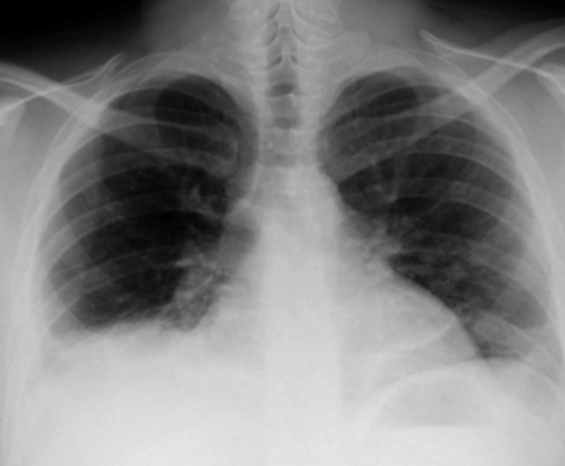
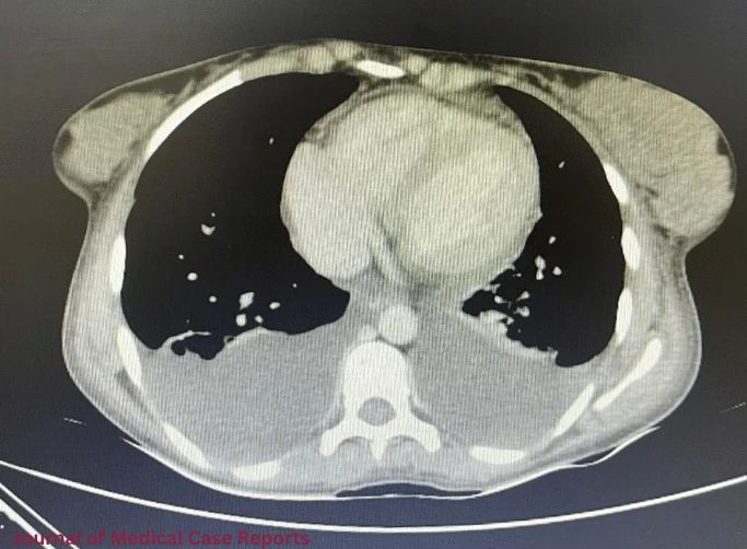
A chest x-ray (CXR) and chest computed tomography (CT) scan showed that both lungs had pleural fluid, which is why both chest tubes were put in. The fluid in the pleura was transudate. A heart ultrasound showed a slight pericardial fluid without endocardial growth.

More tests were done by doctors to figure out how bad the lymphadenopathy was. An ultrasound of the neck showed swollen lymph nodes under the jaw and above the collarbone. The biggest one was 2.5 cm across. A CT scan of the abdomen and pelvis did not show any lymphadenopathy, but it did show hypodense areas in the seventh and sixth segments of the liver.

To find out more, a lymph node biopsy was done with the help of an ultrasound. The results showed that the structure was still there, but there was follicular hypertrophy and necrotizing lymphadenitis with paracortical coagulative necrosis. Immunohistochemistry showed that CD68 and CD4 were expressed, which is consistent with Kikuchi-Fujimoto disease (KFD). A liver core sample from a low-density area also showed limited lobular hepatocyte death and inflammation with histiocytic neutrophils.

Systemic Lupus Erythematosus: How to Treat It

After that, the patient was told that they had KFD-associated systemic lupus erythematosus (SLE) with liver involvement that was not limited to the nodes. Hydroxychloroquine (HCQ) and oral corticosteroids (prednisolone) were used to treat the disease. The patient’s situation improved after five days in the hospital, and she was sent home in good health. After two months of treatment, the lymphadenopathies got better and better until they were gone for good. After six months of regular checks, the symptoms did not come back.

People who have Kikuchi-Fujimoto disease and Systemic Lupus Erythematosus

Kikuchi-Fujimoto disease (KFD), which is also known as histiocytic necrotizing lymphadenitis, is a rare systemic disease that mostly affects young Asian women, though cases have been reported all over the world. KFD can happen to anyone at any age, but most of the time it happens to people under 40.

KFD usually shows up as painful, swollen, and mild necrotizing lymphadenopathy, often with a low-grade fever and tiredness. Headaches, nausea, puking, feeling sick, losing weight, arthralgia, myalgia, night sweats, rashes, and pain in the thorax or abdomen are some of the other symptoms that may happen. KFD can also happen in places other than the nodes, like the skin, bone marrow, and liver, though this is not common. A histological study of lymph nodes usually shows a structure that is only partly intact, with areas of follicular growth and clear areas of necrosis. Different people have different lab readings. Some have average numbers, while others may have anemia, high ESR, CRP, blood LDH, aminotransferases, or leukopenia.

KFD can be mistaken for other infectious, autoimmune, or cancerous diseases because its signs are not always clear. Even though the disease goes away on its own, it is important to get a quick and correct evaluation so that severe treatments are not needed. This study shows a unique case of KFD that affected the liver at the same time as systemic lupus erythematosus (SLE).

Gout and pseudogout in one joint

Seniors often have inflammatory joint illnesses like gout and pseudogout. This case report describes a rare combined occurrence of both illnesses.

A 42-year-old guy complained of right knee discomfort and edema for two days at the emergency room. The pain was spontaneous and trauma-free. For two years, the patient had recurring right knee problems.

The physicians reviewed his right knee arthrocentesis (joint aspiration) findings, which showed negatively birefringent crystals for which he was taking colchicine. Grade 3 right knee chondromalacia, asthma, and ischemic heart disease were mentioned in his medical history.

Examination for Pseudogout and Gout

The physicians found the right knee warm, painful, erythematous, and effusion-prone during a general physical. Also, discomfort limited his mobility. Leukocyte count, erythrocyte sedimentation rate, C-reactive protein, and uric acid were also raised in his labs.

Radiological imaging indicated right knee medial compartment narrowing and degeneration. Joint calcification was not found by radiography. Additionally, joint fluid aspiration showed turbid, straw-colored fluid with high neutrophilic count. Culture and staining were negative, however microscopic examination revealed few positive and negative birefringence crystals. These data supported pseudogout and joint gout diagnoses.

The treatment comprised colchicine and physiotherapy. Due to ischemic heart disease, physicians did not give NSAIDs. Doctors also avoided allopurinol to prevent gout flares. He received paracetamol and tramadol for pain. Patient improved following therapy and released after two days.